

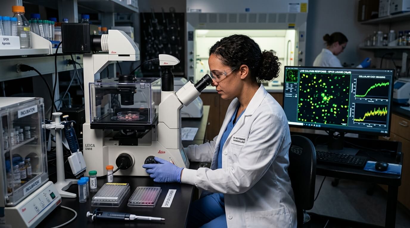
In the world of cell biology, communication is everything. Cells don’t exist in isolation; they are constantly receiving, processing, and responding to signals from their environment. While there are many ways cells send messages, one of the most fundamental and rapid methods involves the movement of calcium ions. This movement, often referred to as calcium signalling, is the reason why the calcium flux assay has become such a cornerstone in modern laboratory research and drug discovery.
Calcium ions ($Ca^{2+}$) act as a universal intracellular messenger. They regulate a vast array of physiological processes, from the way our muscles contract to the way our neurons fire and even how our hearts beat. When a cell is stimulated—perhaps by a hormone, a neurotransmitter, or a potential drug candidate—it often responds by rapidly increasing the concentration of calcium within its cytoplasm. This ‘flux’ of calcium is what researchers are looking for when they want to see if a cell is reacting to a specific stimulus.
How a calcium flux assay actually works in the lab
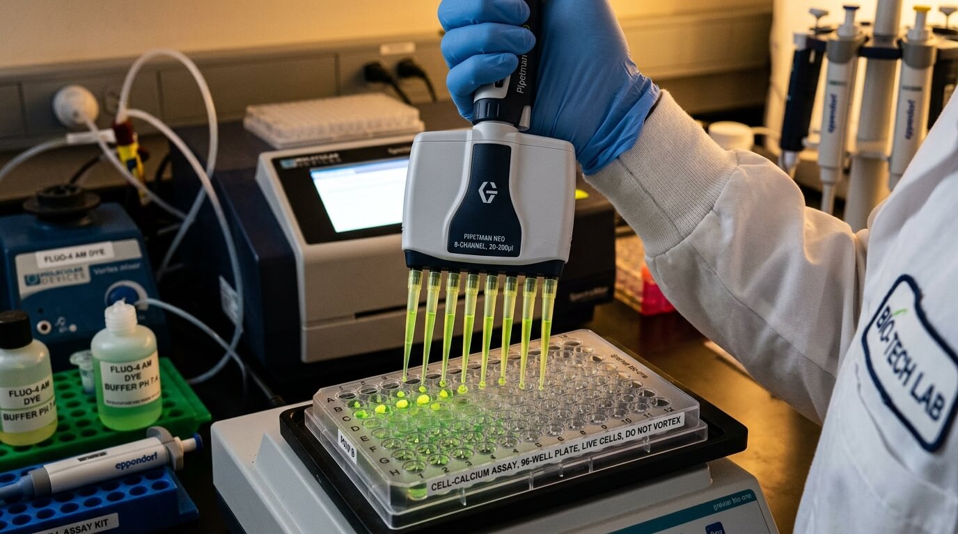
At its heart, a calcium flux assay is designed to monitor the change in intracellular calcium levels in real-time. Because calcium ions themselves are not visible under a standard microscope, scientists use specialised fluorescent dyes to act as indicators. These dyes are engineered to change their light-emitting properties when they bind to calcium ions.
The process generally follows a specific sequence of events to ensure the data collected is both accurate and reproducible. While different laboratories might have slight variations in their protocols, the core steps usually include:
- Cell Preparation: Cells are grown in microplates until they reach the desired density. These could be primary cells or immortalised cell lines specifically engineered to express certain receptors.
- Dye Loading: A calcium-sensitive fluorescent dye is added to the cells. These dyes are often ‘masked’ as esters, allowing them to cross the cell membrane. Once inside, cellular enzymes break down the ester, trapping the active dye within the cell.
- Incubation: The cells are incubated at a specific temperature—often 37°C—to allow the dye to be properly distributed and the cells to recover from the loading process.
- Stimulation and Measurement: The plate is placed into a high-throughput screening instrument, such as a fluorescent plate reader. A stimulus (a ligand or drug) is added, and the instrument measures the resulting change in fluorescence over several seconds or minutes.
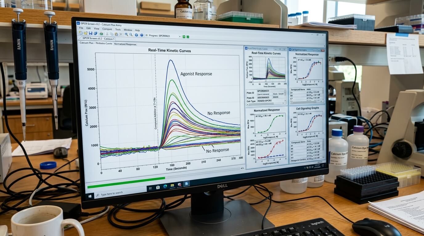
When the stimulus triggers a receptor on the cell surface, it leads to the opening of calcium channels or the release of calcium from internal stores like the endoplasmic reticulum. As the $Ca^{2+}$ concentration rises, more dye molecules bind to the ions, causing a measurable spike in fluorescence. This spike is the ‘flux’ that gives the assay its name.
Why this assay is the gold standard for GPCR research
If you look at the most successful drugs on the market today, a significant portion of them target G-protein coupled receptors (GPCRs). These receptors are involved in almost every aspect of human health, making them the primary focus for pharmaceutical companies. The calcium flux assay is particularly well-suited for studying GPCRs because many of these receptors signal through the $G_q$ pathway, which directly leads to calcium release.
Researchers use these assays to determine several key factors about a potential new drug:
- Agonist Activity: Does the drug activate the receptor and trigger a calcium response?
- Antagonist Activity: Does the drug block a known activator from triggering a response?
- Potency and Efficacy: How much of the drug is needed to achieve a half-maximal response ($EC_{50}$), and what is the maximum response possible?
- Kinetics: How quickly does the response start, and how long does it last?
The ability to gather this data in a high-throughput format means that thousands of compounds can be screened in a single day. This speed is essential in the early stages of drug discovery, where identifying a ‘hit’ compound amongst a library of millions is like finding a needle in a haystack.
The choice between different types of fluorescent dyes
Not all calcium-sensitive dyes are created equal, and the choice of dye can significantly impact the quality of the results. Broadly speaking, researchers choose between ratiometric and non-ratiometric dyes depending on their specific needs and the equipment available in their facility.
Non-ratiometric dyes, such as Fluo-4 or Fluo-8, are popular because they are incredibly bright and easy to use. They show a simple increase in fluorescence intensity as calcium levels rise. However, they can be sensitive to variations in dye loading or cell thickness. If one well in a 384-well plate has slightly more cells than another, the signal might appear stronger even if the calcium flux is the same.
Ratiometric dyes, like Fura-2, work differently. They shift their excitation or emission wavelengths when they bind to calcium. By taking the ratio of the signals at two different wavelengths, researchers can cancel out variables like cell density or dye concentration. This makes ratiometric dyes more precise, though they often require more complex and expensive optical equipment to read.
Optimising the assay for better data quality
While the concept of a calcium flux assay is straightforward, achieving high-quality, reproducible data requires careful optimisation. Small changes in the laboratory environment can lead to significant ‘noise’ in the data, which might mask the subtle effects of a potential drug. Several factors must be meticulously controlled to ensure the assay remains robust.
Temperature control is perhaps one of the most overlooked aspects. Many ion channels and receptors are highly temperature-sensitive. If the assay is performed at room temperature rather than at physiological 37°C, the kinetics of the calcium movement might be slowed down, leading to inaccurate conclusions about how a drug might behave in the human body. Similarly, the pH of the buffer used during the assay must be tightly regulated, as fluctuations can interfere with both the health of the cells and the performance of the fluorescent dye.
Another common challenge is ‘background’ fluorescence. Some chemical compounds in a drug library might naturally be fluorescent themselves, which can lead to false positives. To combat this, researchers often use ‘no-wash’ assay kits that include masking dyes. These masking dyes stay outside the cells and quench any fluorescence that isn’t coming from the intracellular calcium indicators, significantly improving the signal-to-noise ratio.
The move towards more complex cell models
In recent years, there has been a shift away from using simple, engineered cell lines towards more biologically relevant models. While immortalised cells are great for consistency, they don’t always reflect the complexities of human physiology. This has led to the increased use of primary cells and induced pluripotent stem cell (iPSC)-derived cells in calcium flux assays.
For example, iPSC-derived cardiomyocytes (heart cells) are now frequently used to test for potential drug toxicity. Because these cells naturally beat and exhibit regular calcium oscillations, researchers can use a calcium flux assay to see if a new drug disrupts these rhythms. This is a critical step in ensuring that a new medication won’t cause dangerous side effects like arrhythmias before it ever reaches human clinical trials.
The versatility of the assay also extends into neurobiology. By monitoring calcium flux in neurons, scientists can study synaptic activity and the way different neurotransmitters influence brain health. Whether it is looking for treatments for Alzheimer’s disease or chronic pain, the ability to see ‘live’ cellular responses provides a level of insight that static assays simply cannot match.
Modern instrumentation and high-throughput capabilities
The evolution of plate readers has been a game-changer for this field. Modern systems are now capable of reading 384-well or even 1536-well plates simultaneously. These instruments are equipped with integrated fluidics, meaning they can add the stimulus and begin recording the fluorescence change in a fraction of a second. This is vital for capturing the ‘peak’ of the calcium flux, which often happens very rapidly after stimulation.
Beyond just measuring intensity, some advanced systems can now perform imaging-based calcium assays. This allows researchers to see not just that the calcium level in a well has increased, but exactly which cells within that well are responding. This spatial information can be incredibly valuable when working with mixed cell populations or complex tissue models where different cell types might respond differently to the same stimulus.

James provides insightful news coverage, practical tips, and informative articles to keep readers updated and engaged.